What exactly is an Echocardiogram?
An Echocardiogram, also known as an echo.
It is a type of ultrasound examination that uses sound waves to create moving
images of your heart.
What is the purpose of an Echo-Cardiogram?
An Echo-Cardiogram can assist your doctor in learning vital information about your heart health. It will determine whether your heart, heart valves, and the amount of blood pumped out by your heart are normal or if you have heart disease or another heart condition.
-
If you have any of the following symptoms, you may require an
echo-cardiogram:
- Chest pain or pressure,
- Shortness of breath,
- Irregular heartbeats,
- Abnormal heart sounds are all signs and symptoms of a heart problem.
- Have you had a heart attack or had heart surgery?
- Have a heart condition such as cardiomyopathy (heart muscle disease) or heart valve disease?
What Should You Expect During an Echocardiogram?
A specially trained technician will perform your test in a doctor's
office or diagnostic center,or hospital. There are no special preparations required, and
the test usually lasts 30 to 60 minutes.
The test will be performed in a darkened room so that your technologist can see the test
monitor more clearly. You will undress from the waist up and put on a hospital gown
before lying down on an exam table.
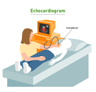
Your technologist will stick electrodes to your chest to monitor your heart rhythm with
an electrocardiogram or EKG. Next, your technologist will apply gel to your chest and
move a small device called a transducer back and forth against your chest over your
heart. The transducer detects and transmits sound waves, which travel to the test
monitor and display images of your heart.
You will spend the majority of the test lying on your left side. It is possible that you
will need to lie on your back and hold your breath for a few seconds at a time. You may
hear a whooshing sound during the test, which is the sound of blood moving through your
heart.
The Results of an Echocardiogram
In most cases, your doctor will have the results of your
echo-cardiogram within 30 minutes to 1 Hour and will go over them with you. The
following outcomes are possible:
Normally refers to your heart, heart valves, and the amount of blood your heart pumps
out.
Abnormal, which may include your heart chambers or valves not working properly, the
amount of blood your heart pumps out being insufficient to meet your body's needs, there
being extra fluid around your heart, or you having a tumour or blood clot in your heart.